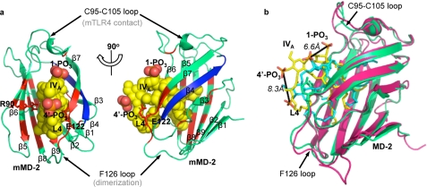
FIGURE 2.
Lipid IVA packs tilted and shallowly in the mouse MD-2 hydrophobic pocket in the docked mMD-2·lipid IVA complex. The docked mMD-2·lipid IVA complex is shown in two perpendicular views in a and overlaid to the hMD-2·lipid IVA co-crystal structure in b. MD-2 is shown in ribbon views in a and b, and lipid IVA is shown in sphere view in a and stick view in b. The residues interacting with lipid IVA are colored red, and the β4 strand is colored blue in a. The fourth acyl chain of lipid IVA is labeled L4 in both a and b. Green, mMD-2; yellow, lipid IVA in mMD-2; pink, hMD-2; cyan, lipid IVA in hMD-2. The graphics were created using PyMol (DeLano Scientific).

