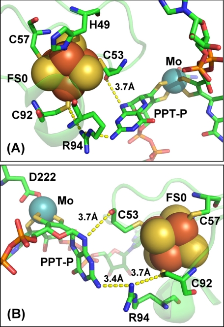
FIGURE 1.
Structure and potential electron transfer routes between FS0 and the Mo-bisPGD cofactor of NarGHI. Two views of the FS0- and Mo-bisPGD-coordinating region of NarG are shown, with a rotation on the vertical axis of ∼180° between the two panels (A and B). Shown in the figure is the position of the two residues mutated in this work (NarG-His49 and NarG-Arg94), as well as putative electron transfer pathways between FS0 and the proximal pyranopterin (PPT-P). Distances within these putative pathways are indicated in the figure. In this figure, iron, sulfur, molybdenum, oxygen, carbon, and phosphorus atoms are rendered in red, yellow, blue, red, green, and orange, respectively.

