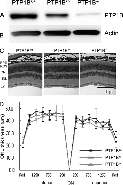
FIGURE 5.
Morphological analysis of PTP1B knock-out mice born and raised in dim cyclic light. Retinal proteins from wild-type, heterozygous, and homozygous PTP1B knock-out mice were subjected to immunoblot analysis with anti-PTP1B (A) and anti-actin (B) antibodies. C, hematoxylin and eosin-stained retinal sections of the retina from the eyes of wild-type, heterozygous, and homozygous PTP1B mice at 6–8 weeks of age. D, quantification of morphological changes. Plots of total retinal thickness in the inferior and superior regions of the retinas of wild-type, heterozygous, and homozygous PTP1B mice are shown. Values are mean (S.D.), n = 2 (wild-type), 3 (heterozygous), and 3 (homozygous). Examination of retinas from each group revealed no structural differences in any of the retinal cells at the light microscope level. RPE, retinal pigment epithelium; ROS, rod outer segments; ONL, outer nuclear layer; INL, inner nuclear layer; GCL, ganglion cell layer.

