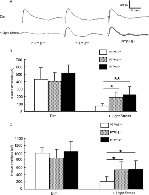
FIGURE 7.
Functional assay of PTP1B knock-out mice after light damage. The ERG amplitudes were recorded for wild-type, heterozygous, and homozygous PTP1B knock-out mice after light exposure at 14,000 lux for 7 days. A, representative ERG tracing from wild-type, heterozygous, and homozygous PTP1B knock-out mice before and after light stress. Average a- (B) and b-wave (C) amplitudes of scotopic ERG were measured from wild-type, heterozygous, and homozygous PTP1B knock-out mice. The a-wave amplitude was measured from the resting level to the peak of the cornea-negative deflection, and the b-wave amplitude was measured from the trough of the a-wave to the crest of the cornea-positive response. Data are mean (S.D.), and the number of animals used in this study were 6 (wild-type), 6 (heterozygous), and 5 (homozygous). *, p < 0.05 and **, p < 0.01 by one-way analysis of variance followed by Scheffe's posthoc test.

