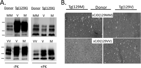
FIGURE 1.
Transmission of sCJD to Tg(129V) and Tg(129M) mice. A, Western blots of rPrPSc from brain homogenates of the original human sCJD(129MM) and sCJD(129VV) donors and a representative Tg(129V) or Tg(129M) recipient mouse. Samples were freshly prepared from frozen frontal cortex as 10% (w/v) brain homogenate in lysis buffer. Total protein was normalized and subjected to 20 μg/ml of PK for 30 min at 37 °C, probed with monoclonal antibody 3F4. Markers on the left represent 37, 25, and 20 kDa, top to bottom. All mice were clinically sick, as defined under “Materials and Methods.” B, spongiform degeneration evident in hematoxylin and eosin (H&E) stainings of sections of the frontal cortex taken from sick Tg(129M) (left sections) and Tg(129V) (right sections) mice inoculated with brain homogenate from sCJD(129MM) (top sections) or sCJD(129VV) (bottom sections), as indicated. PrP plaques were not present in any brain region.

