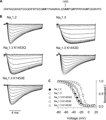
FIGURE 7.
A, amino acid sequence of the Nav1.3 channel III-IV linker region is shown, with lysine 1453 indicated. The sequence of the Nav1.2 III-IV linker region is identical. B, sample current traces for Nav1.3 lysine 1453 mutants during the 5-mV test depolarization of the inactivation protocol, as described under “Experimental Procedures.” C, voltage dependence of inactivation is shown for Nav1.3 K1453Q (triangles), Nav1.3 K1453E (squares), and Nav1.3 K1453D (diamonds) mutants and wild type Nav1.2 (black circles) and Nav1.3 (white circles) channels. The parameters of the fits and sample sizes are shown in Table 4. Data points indicate means, and error bars show S.D. values.

