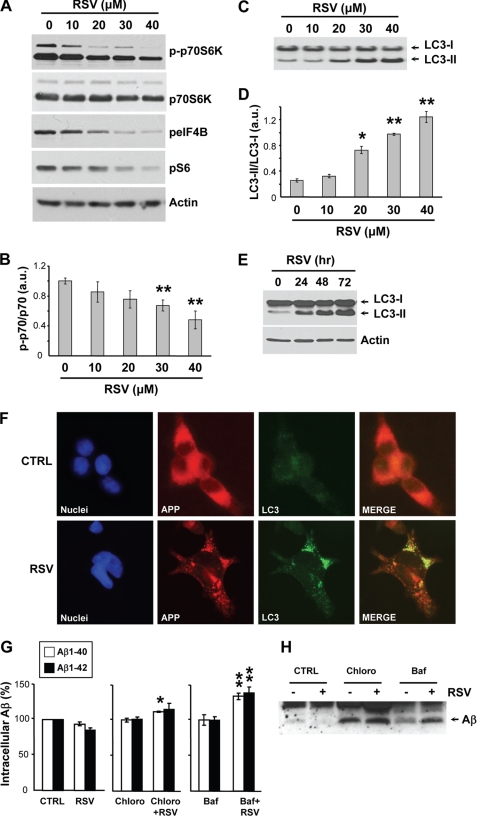
FIGURE 5.
Resveratrol induces autophagy and lysosomal degradation of Aβ. A–D, APP-HEK293 cells were treated for 24 h with the indicated concentrations of resveratrol (RSV). Cell extracts were then analyzed by WB for phospho-p70S6K (p-p70S6K), p70S6K, phospho-eIF4B (peIF4B), phospho-S6 (pS6), and actin (A) and for LC3 (C). Densitometric analysis and quantification of the p-p70S6K/p70S6K (B) and LC3-II/LC3-I (D) ratios are shown. a.u., arbitrary units. E, WB analyses of LC3 and actin in APP-HEK293 cells treated for the indicated times with 40 μm RSV. F, immunocytochemistry with anti-APP (red) and anti-LC3 (green) antibodies in APP-HEK293 cells incubated with for 72 h in the absence (CTRL) or presence of 40 μm RSV. Nuclei were stained with 4′,6-diamidino-2-phenylindole (DAPI, blue). G and H, shown are ELISA measurements of intracellular Aβ1–40 and Aβ1–42 levels (G) and WB analysis of immunoprecipitated intracellular Aβ (H) from APP-HEK293 cells treated for 24 h with 40 μm RSV or its vehicle (DMSO, CTRL) in the absence or presence of chloroquine (Chloro, 100 μm) or bafilomycin A1 (Baf, 100 nm). Histograms in B, D, and G show the mean ± S.D. of three independent experiments. *, p < 0.05; **, p < 0.01 (Student's t test).

