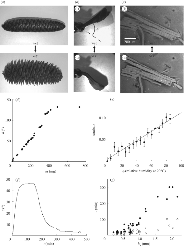
Figure 1.
(a) Cone from Picea abies in its wet (closed) and dry (open) states. (b) Cone scale in its wet (i) and dry (ii) states. We call θ the angular position of the scale, the dry state being chosen as a reference (θ = 0°). (c) Environmental scanning electron microscopy (ESEM) images of a few cells from the responsive tissue of a scale of Pinus coulteri. The cells are about 20 per cent longer in the wet state (i) than in a dry environment (ii). (d) Opening angle of a scale of P. coulteri as a function of its water content. The reference of angle is chosen for a dry sample. (e) Single-cell ESEM measurements of strain–humidity relationship in the active tissue of a scale of P. coulteri. Strains are measured along the axis of the cells, the radial expansion being negligible. The line is a linear fit to the data with equation ε = 0.0011ϕ + 0.0033. (f) Plot of the opening angle θ of a scale versus time, when immersed in water and then drying in ambient air. The opened (dry) position was chosen to be the zero angle reference. (g) Opening and closing times of scales of different sizes as a function of the thickness ha of external (i.e. responsive) layer of cells. Filled circles are for opening times (in a 20°C and 40% humidity environment) and open circles for closing times.

