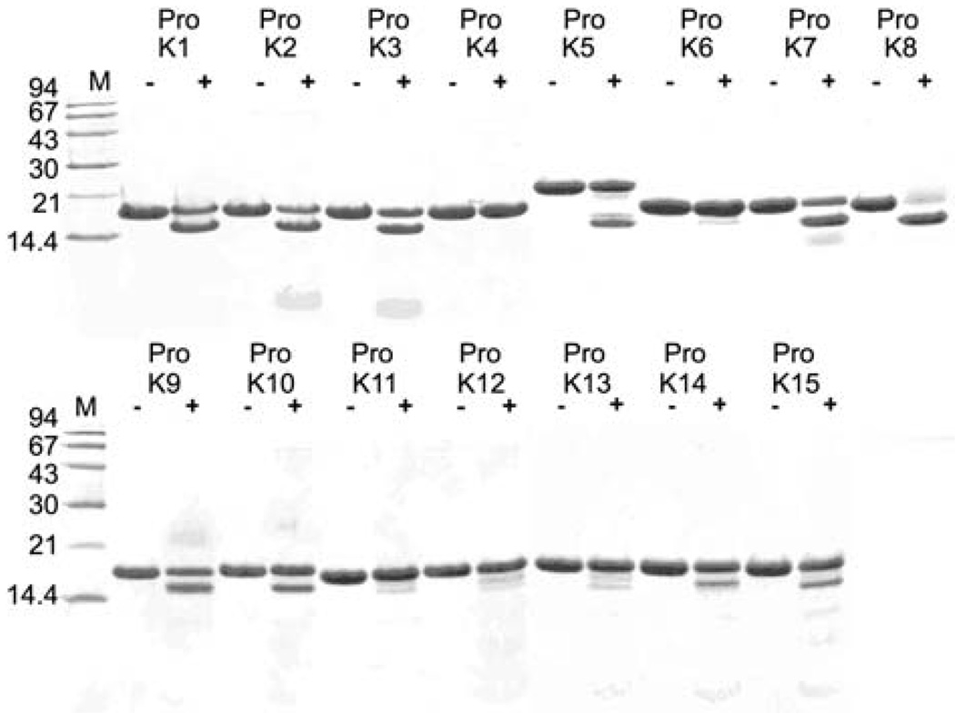
Figure 1. Gel electrophoretic analysis of pro-KLK1–15 fusion protein cleavage.
Coomassie Brilliant Blue-stained 16.5% Tricine sodium dodecyl sulfate-polyacrylamide gel after separation of the products of 100:1 molar ratio incubation of pro-KLK1–15 fusion proteins (abbreviated as ‘Pro K’; final concentration of 40 µm) with KLK15 for 24 h at pH 7.4.

