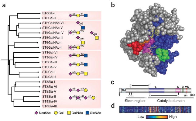
Figure 1.
Structural relationships among the human sialyltransferase family. a) Homology dendrogram is shown for twenty members of the human siatlyltransferase family. Sialoside products produced by each of the four major subfamilies are shown in symbol form. b) CPK representation of pST3Gal-I highlighting the four ‘sialyl motifs’: large (L, cyan), small (S, red), 3 (3, green) and very small (VS, yellow). The bound acceptor ligand is highlighted in red. The postulated ‘lid’ domain consisting of a disordered loop located between the motifs 3 and VS is omitted. c) Localization of the sialylmotif regions in the primary sequence of pST3GalI highlighted using the same color-coding for the conserved motifs in panel b. d) Homology heat map highlighting regions of sequence conservation among all 20 members of the sialyltransferase family. relative to hST3GalI. Insertions in other sialyltransferases relative to hST3Gal I are omitted to maintain comparisons relative the sialylmotifs illustrated in panel c.

