
Figure 3.
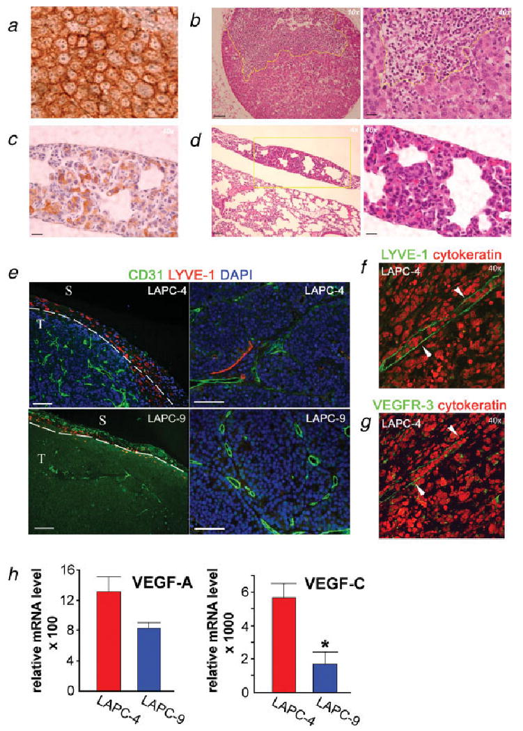
Immunohistochemical detection of metastases and studies of angiogenic and lymphangiogenic tumor profiles. Lung and lymph node sections from LAPC-4- and LAPC-9-implanted mice were processed to detect tumor cells in the parenchyma. LAPC-4 primary tumor sections served as positive controls for human cytokeratin (a). Hematoxylin & eosin stained sections revealed tumor metastases in the lymph nodes (b) and lungs (d) from the LAPC-4 group. A brachial lymph node from a LAPC-4-implanted mouse shows a large subcapsular lesion (b). Cytokeratin staining revealed tumor cells present in lungs in the LAPC-4-implanted group (c). Yellow lines indicate the tumor mass. Scale bars = 200 μm (×4), 100 μm (×10) and 25 μm (×40), respectively. The blood (CD31; green) and lymph (LYVE-1; red) vessels of the primary tumors (nuclei, blue) were visualized by immunohistochemistry. Analysis of LAPC-4 tumors revealed the presence of small lymphatics both in peritumoral and intratumoral areas (e, left: ×10 and right: ×20; f, ×40). Most LAPC-9 tumors displayed only very few lymphatic vessels at the tumor periphery (e, left: ×10). Scale bar in e = 100 μm. T, tumor; S, surrounding tissue. The intratumoral LYVE-1 positive (green) structures (f, ×40, white arrowheads) were also positive for another lymphatic marker, VEGFR-3 (green) (g, ×40, white arrowheads). Angiogenic and lymphangiogenic growth factor expressions were analyzed in the excised tumors by real time RT-PCR analysis (h). VEGF-A levels were found to be similar whereas VEGF-C expression levels were significantly and markedly elevated in LAPC-4 as compared to LAPC-9 tumors. *p < 0.05.