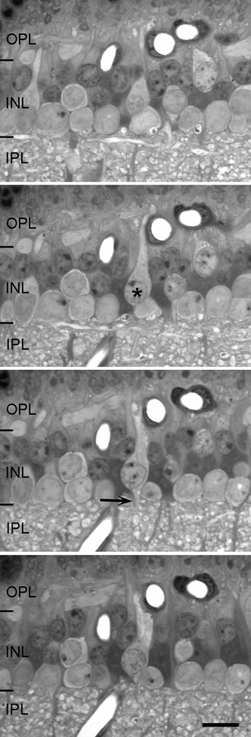
Figure 2.
Four sequential sections, each 1 µm in thickness, of the inner plexiform layer. By looking at consecutive serial sections, it is possible to identify each cell by studying the location of its cell body, its staining characteristics and the termination patterns of its processes. This particular example shows a bipolar cell (asterisk) in vertical section with dendrites reaching toward the outer plexiform layer (OPL) and a thin axon (arrow) exiting from the cell’s soma into the inner plexiform layer (IPL). Scale = 10 µm.

