
FIGURE 2. Detection of transient conserved intermediate species during CsgA polymerization.
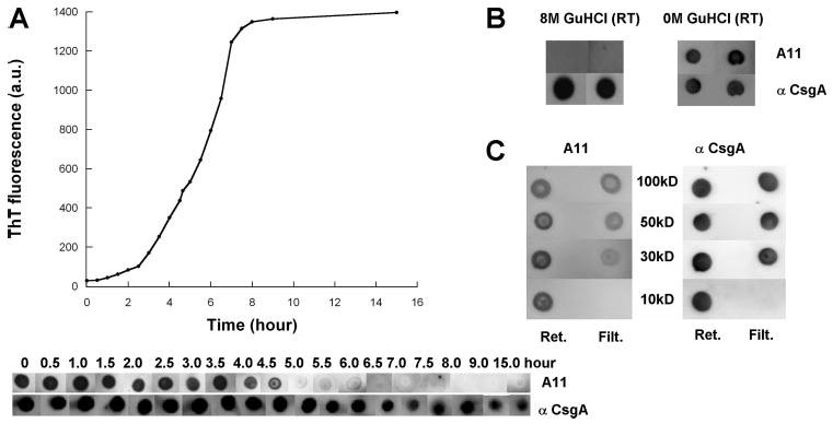
A, ThT fluorescence (top) and immunoblotting (bottom) of 80 μm CsgA incubated for the indicated time after purification. At the indicated times, samples were removed, ThT was added to a final concentration of 20 μm, and fluorescence was measured. Samples were blotted onto a nitrocellulose membrane and probed with the A11 antibody, and after stripping, probed with the anti-CsgA antibody. a.u., arbitrary units. B, CsgA denatured with 8 M GdnHCl was blotted onto nitrocellulose and probed with the A11 and anti-CsgA antibodies (left). GdnHCl (GuHCl) was removed using a Sephadex G25 column (final buffer: 50 mm KPi, pH 7.2) and then immediately blotted onto nitrocellulose and probed with the A11 and anti-CsgA antibodies (right). RT, room temperature; Ret., retentate; Filt., filtrate. C, Amicon ultra filters were used to separate CsgA solutions prior to probing with the A11 and anti-CsgA antibodies. The molecular weight cutoff of the filters is indicated. Retentates and filtrates were immediately blotted onto nitrocellulose and probed with the A11 antibody, and after stripping, probed with the anti-CsgA antibody.