Fig. 6.
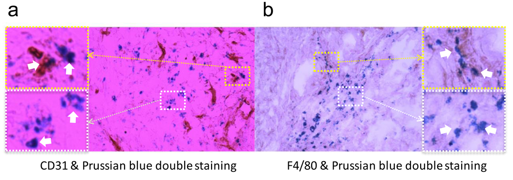
(a) CD31 and Prussian blue double staining of the tumor samples. Some particles were found within the vessels (upper left), while many others managed to extravasate (lower left). (b) F4/80 and Prussian blue double staining of the tumor samples. Although some particles were found within macrophages (upper right), most of them were found independent of macrophages (lower right).

