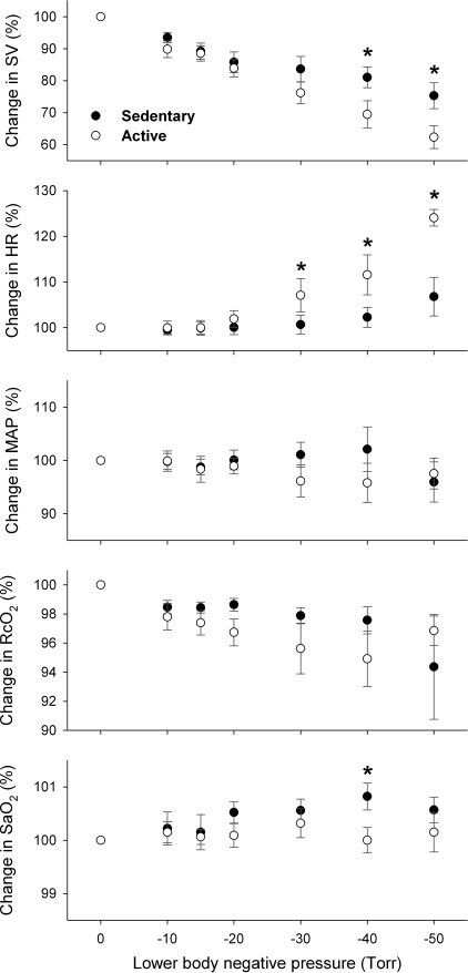
Fig. 2.
Changes in stroke volume (SV), heart rate (HR), MAP, regional cerebral tissue oxygenation (RcO2), and systemic arterial O2 saturation (SaO2) during lower body negative pressure (LBNP). Graded decreases in SV were associated with progressive increases in LBNP (P = 0.0001), suggesting central hypovolemia in response to LBNP-simulated orthostatic challenge. This decrease in SV appeared more substantial in the ACT group than in the SED group (P = 0.0006). *Significant difference between the groups. A tachycardiac response occurred from a LBNP of −30 Torr (P = 0.0001). This baroreflex-mediated HR was significantly more sensitive in the ACT group in than the SED group (P = 0.0003). MAP was similarly maintained during LBNP in both fitness groups (P = 0.89). RcO2 was diminished during LBNP (P = 0.0187), which was not significantly different between the groups (P = 0.19). The change in SaO2 was not significantly altered by LBNP (P = 0.35), which appeared to become higher in the SED group compared with the ACT group (P = 0.0241).

