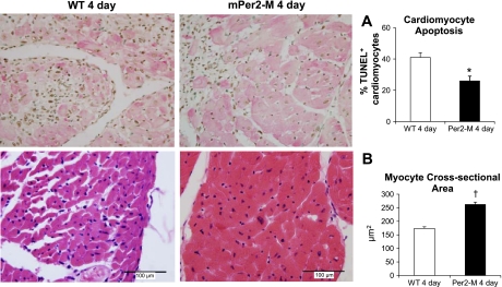
Fig. 2.
Right: reduced cardiomyocyte apoptosis (A) and increased myocyte cross-sectional area (B) in mPer2-M mouse hearts 4 days after chronic infarction are shown. Left: there were 15% less terminal deoxynucleotide transferase-mediated dUTP nick end labeling (TUNEL)-positive apoptotic cardiomyocytes observed in mPer2-M mice (top, right) compared with WT (top, left; *P < 0.05). The average myocyte cross-sectional area (in μm2; †P < 0.001) was increased in mPer2-M mice (bottom, right) vs. WT (bottom, left).

