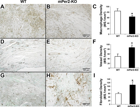
Fig. 3.
Pan-leukocyte marker CD45 staining showed reduced CD45-positive inflammatory cells in mPer2-M mice (A) compared with WT mice (B) at 4 days after infarction (C; *P < 0.05). KO, knockout. There was a significant increase in vessel density in mPer2-M (E) hearts compared with WT hearts (D) at 4 days post-MI (F; †P < 0.01). Fibroblast density was also increased in mPer2-M (H) compared with WT (G) mouse hearts at 4 days post-MI (I; *P < 0.05).

