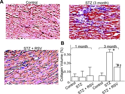
Fig. 2.
A: representative micrographs (×40) of formalin-fixed, paraffin-embedded sections obtained from LV apex of control and 3 mo untreated (STZ) and RSV-treated (STZ + RSV) diabetic animals. Sections (5 μm) were stained with trichrome-masson and counterstained with hematoxylin and eosin (H & E). B: collagen volume fraction (CVF) analysis in different groups of animals after 1 and 3 mo experimental period. CVF was calculated by measuring the area of blue-stained collagen fibers in the interstitial space and expressed as %total area examined. A minimum of 10 different areas were examined in four different hearts of each group. P < 0.05 compared with *control group and †STZ group.

