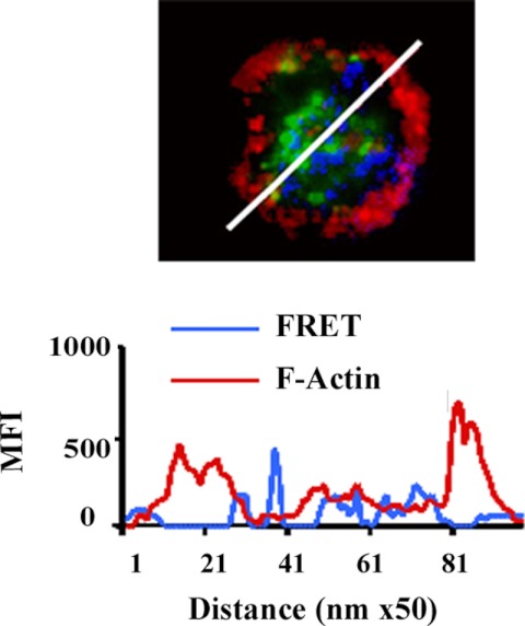
Fig. 5.
Cellular localization of IL-18 association with F-actin. In the micrograph IL-18 (green) does not appear to associate with the dense cortical F-actin (red) in resting neutrophils, as assessed by the lack of a FRET+ between F-actin and IL-18 (blue) in the cellular periphery. To better demonstrate the locality of IL-18 and dense cortical actin, a line scan was performed through the cell (white line), and below it is a graphic representation of the pixels per nanometer of the FRET between IL-18 and F-actin (blue) demonstrating that this interaction appeared in the cytosol and did not localize with the dense cortical F-actin immunoreactivity (red) present at the cell periphery. Multiple random diameters were examined (n = 9) on PMNs from at least 3 donors with similar results and FRET efficiencies of 32–33% MFI, mean fluorescence intensity. Figure is representative of 3 identical experiments.

