Fig. 1.
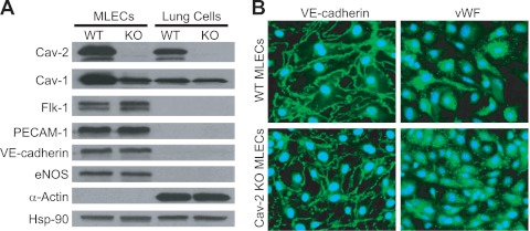
Isolation and initial characterization of mouse lung endothelial cells (MLECs). A: Western blot characterization of the purity of isolated wild-type (WT) and caveolin-2 (Cav-2) knockout (KO) MLECs relative to “lung cells” remaining after first immunoisolation with anti-platelet endothelial cell adhesion molecule-1 (PECAM-1) antibody. Note that, unlike WT, Cav-2 KO MLECs and lung cells are completely devoid of Cav-2. Both WT and Cav-2 KO MLEC protein lysates are positive for endothelial cell (EC)-specific marker proteins, i.e., Flk-1, PECAM-1, VE-cadherin, and endothelial nitric oxide synthase (eNOS), but negative for the mural cell marker protein α-actin. Conversely, protein lysates from lung cells are negative for EC marker proteins but positive for α-actin. B: immunofluorescent microscopy images of MLECs colabeled with 4′-6-diamidino-2-phenylindole (DAPI; blue nuclear staining) and with antibodies against EC marker proteins: VE-cadherin (green plasma membrane staining) or von Willebrand factor (vWF; green perinuclear staining). Note that all WT and Cav-2 KO cells are VE-cadherin or vWF positive. Hsp-90, heat shock protein 90.

