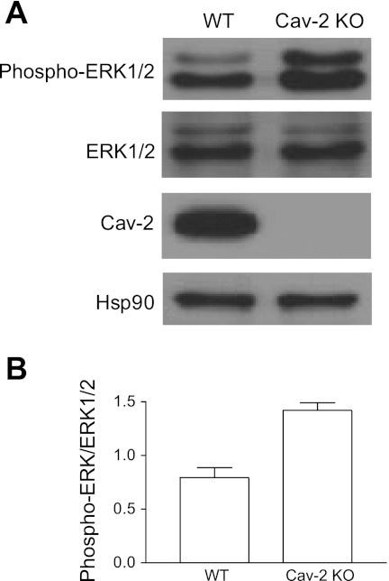
Fig. 7.
Augmented activation/phosphorylation state of ERK1/2 in hyperproliferating Cav-2 KO relative to WT MLECs determined by immunoblotting. A: WT and Cav-2 KO MLECs were plated at 3 × 10⁵ per 150-mm dish, and 2 days later, were lysed and immunoblotted with the indicated antibodies. B: mean densitometric ratios ± SD (n = 3) of phosphorylated ERK1/2 (phospho-ERK1/2) to total expression level (ERK1/2).

