
Fig. 1.
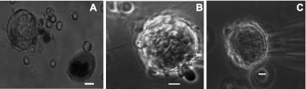
Sample megakaryocytes (MKs) studied on the basis of morphology. Brightfield photomicrographs (×40). A: stage I MK illustrating a viable cell on the left and a darker nonviable cell on the right (Trypan blue exclusion viability test). Phase contrast photomicrographs (×60). B: stage II MK with protruding pseudopodia. C: stage III MK ruffling and blebbing. Cells were in extracellular recording solution (ES) at room temperature. Calibration bars represent 10 μm.