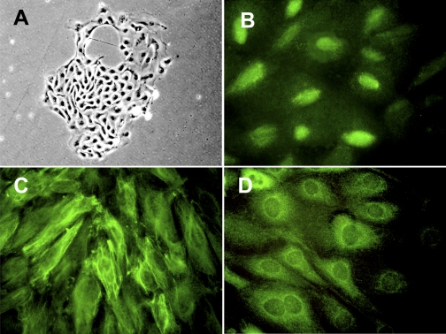
Fig. 1.
Proliferating primary urinary cells display podocyte markers. Urinary cells from a healthy volunteer were cultured in growth medium on a type I collagen-coated dish. Colony formation observed at 2 wk after start of culture demonstrated a cobblestone appearance under phase-contrast microscopy (A). Immunofluorescent staining identified Wilms' tumor 1 (WT1) in nuclei (B), nestin in cytoplasm with a filamentous pattern (C), and synaptopodin in cytoplasm with a partially filamentous and partially homogenous pattern (D) in the cells. Original magnification: ×200 (A); ×400 (B–D).

