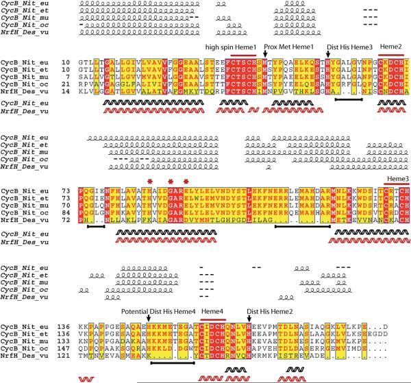
Figure 2.
Clustal-W multiple-sequence alignment of NrfH from D.vulgaris and CycB proteins from ammonia oxidizers. Predicted secondary structure (helical) elements are shown above the alignment, while helices below represent elements based on the crystal structure (NrfH) and homology modeling (cyt cm552). Alignment analyzed using ESPript (Easy Sequencing in PostScript) visualization. A white letter with a red background designates strict identity; a red letter designates similarity in a group, and a yellow background designates similarity across groups. Arrows indicate probable axial ligands in CycB proteins based on alignment to NrfH (see the text). Asterisks indicate aligned ligands proposed to be involved in Q binding in NrfH (see the text). Black lines below the alignment represent homologous regions in CycB not present in NrfH.

