
FIG. 3.
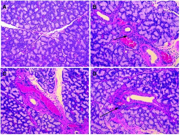
Histopathology with H&E stain sLG. (A) Superior LG (sLG) sections from normal sLG was similar to iLG. (B) sLG from the ID group showed infiltrating cells around ducts and venules. (C) The ID/E group showed infiltration but not as common as seen in ID group. (D) Sections of sLG from ID/Rx rabbits rarely displayed immune cells compared to ID animals.