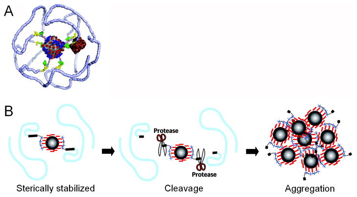
Figure 4.
Protease-sensitive agents for MRI. A) The agent consists of a 5 nm magnetite core (gray) covered by a negatively charged citrate shell (red). The peptide-mPEG copolymers are electrostatically bound by the positively charged coupling domains (blue). The mPEG polymers (light blue) are linked to the coupling domain via the cleavage domain (yellow) and a linker peptide. A model of MMP-9 (brown) is shown for size comparison at the cleavage domain. Green: fluorescein dyes. B) When the sterically stabilized agent is exposed to a protease, the peptide-mPEG linkage is cut at the cleavage domain, resulting in a loss of sterical stabilization. This leads to particle aggregation and enhanced MR signal. Adapted from 76.

