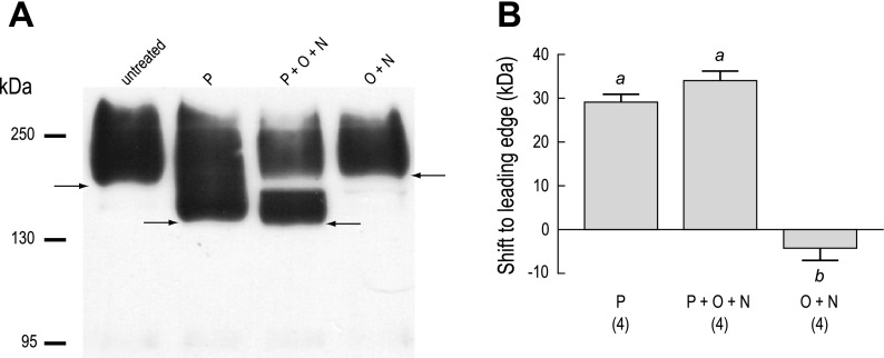
Fig. 6.
Glycosylation of heterologously expressed AeAE in Xenopus oocytes. A: Western blot of total membrane fractions isolated from AeAE oocytes (6 days postinjection). The membrane fractions were either untreated or exposed to one of the following enzymatic treatments: 1) PNGase F (P); 2) a mixture of PNGase F, O-glycosidase, and neuraminidase (P + O + N); or 3) a mixture of O-glycosidase and neuraminidase (O + N). The anti-AENt antibody was used to detect AeAE immunoreactivity. Arrows indicate the leading edge of the immunoreactivity. Migrations of the molecular mass markers (in kDa) are indicated at left. B: summary of the effects of enzymatic deglycosylation on the migration of the AeAE immunoreactivity. Shaded bars represent the shift (in kDa) to the leading edge. Values are means ± SE based on the number of measurements shown in parentheses. a,bP < 0.001, categorization of the means as determined by a repeated-measures ANOVA and Newman-Keuls posttest.

