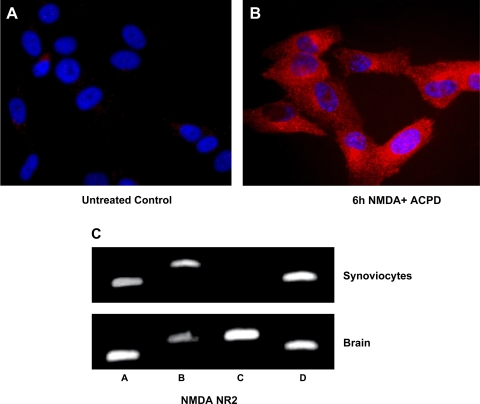
Fig. 4.
Glutamate NMDA NR2 receptor subunit expression on synoviocytes identified by immunostaining and RT-PCR. A: high-power photomicrograph of glutamate NMDA NR2 receptors (red) on control untreated clonal SW892 human synoviocytes synovial cells. B: immunostaining was increased in cells incubated concurrently with 5 μM NMDA and 5 μM ACPD for 6 h. Cells shown were stained with rabbit anti NMDA NR2 antibody. Bar, 20 μm. C: amplification of specific NMDA NR2 subunits was identified by RT-PCR in human synoviocytes. Cytosolic lysates of untreated SW982 cells (synoviocytes) and human brain (hippocampus) as control were applied to SDS-PAGE and transferred to a nitrocellulose filter. Lanes were primed for NMDA NR2 subunits A–D as shown. All 4 subunits were expressed in the lysates from human brain. NMDA NR2 subunits A, B, and D were expressed in lysates from untreated SW982 synoviocytes.

