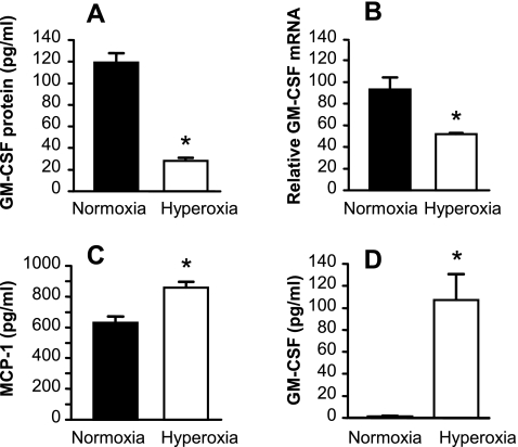
Fig. 1.
Effects of exposure to hyperoxia on lung epithelial cell expression of granulocyte/macrophage colony-stimulating factor (GM-CSF) and MCP-1. Primary murine alveolar epithelial cells (AEC) (A–C) or MLE-12 cells (D) were exposed to hyperoxia (80% oxygen/5% CO2) or normoxia (21% oxygen/5% CO2) for 48 h. The culture medium was then replaced with fresh medium, and the cells returned to hyperoxic or normoxic conditions for 18 h further for determination of GM-CSF and MCP-1 protein in the culture supernatant. All data are expressed as means ± SD, n = 3, and are representative of 3 independent experiments involving separate type II cell isolations. A: GM-CSF protein in culture supernatants from AEC. *P < 0.0001. B: relative GM-CSF mRNA in primary AEC was measured by real-time PCR. *P < 0.002. C: MCP-1 protein in culture supernatants from primary AEC. *P < 0.002. D: GM-CSF protein in culture supernatants from MLE-12 cells. *P < 0.001.

