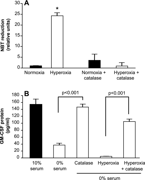
Fig. 6.
Effects of catalase on AEC oxidative stress and GM-CSF expression following exposure to hyperoxia. Primary murine AEC were treated with PEG-catalase (1,000 U/ml) during exposure to hyperoxia or serum deprivation. Control AEC were maintained in 10% serum in normoxia. Data are expressed as means ± SD. The experiments shown are representative of 3 independent experiments. A: AEC ROS were measured as NBT reduction; n = 5. *P < 0.001 vs. each of the other conditions. There was no significant difference between normoxia, normoxia + catalase, and hyperoxia + catalase. B: GM-CSF protein was measured in culture supernatants. Data are expressed as means ± SD. P < 0.001 comparing catalase vs. no catalase, n = 3.

