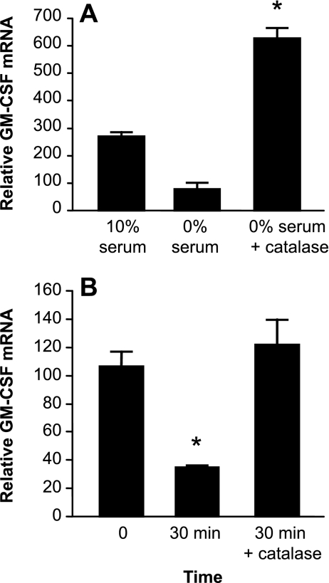
Fig. 7.
Effect of catalase on GM-CSF mRNA expression and stability during oxidative stress. Primary murine AEC were exposed to serum deprivation with or without the addition of PEG-catalase (1,000 U/ml). Additional control cells were cultured in 10% serum. A: relative GM-CSF mRNA expression in cells exposed to serum deprivation. Data are expressed as means ± SE, n = 3. *P < 0.001 vs. 0% serum. B: after 24 h of oxidative stress, AEC were stimulated with IL-1β for 3 h before the addition of Actinomycin D (5 μg/ml) to stop transcription. Relative GM-CSF mRNA was determined by real-time PCR at 0 and 30 min. Data are expressed as means ± SE, n = 4. *P < 0.01.

