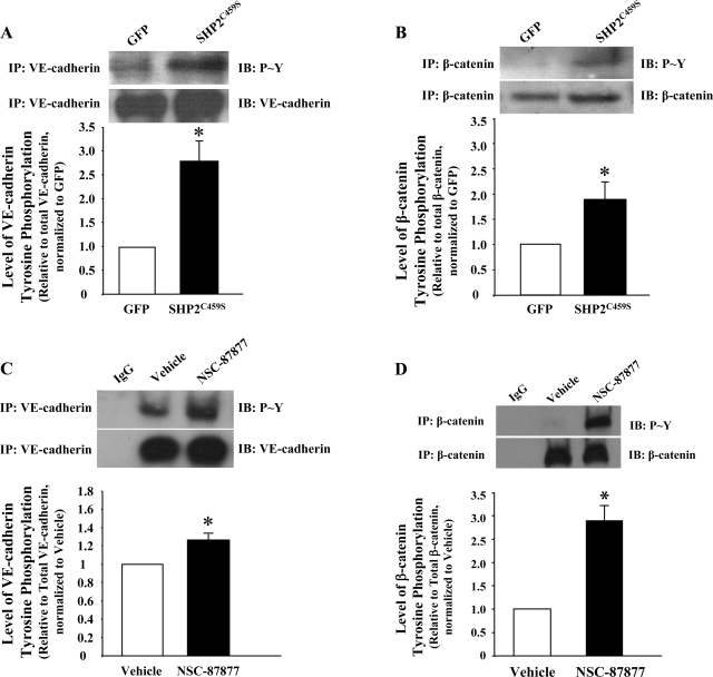
Fig. 3.
Inhibition of SHP2 leads to increased tyrosine phosphorylation of adherens junction proteins. Equivalent numbers of PAEC were transfected with GFP or SHP2C459S for 48 h (A and B) or confluent monolayers of lung microvascular endothelial cells (LMVEC) were incubated with 100 μM NSC-87877 for 3 h (C and D). Equal amounts of endothelial cell lysate were immunoprecipitated with antibodies directed against VE-cadherin (A and C) or β-catenin (B and D), and precipitates were immunoblotted for phosphorylated tyrosine. The immunoblots were stripped and reprobed for the immunoprecipitated protein. The data are presented as means ± SE of densitometric values of tyrosine phosphorylated adherens junction protein relative to total adherens junction protein, normalized to GFP (A and B) or vehicle (C and D). N = 3; *P < 0.05 vs. GFP or vehicle, respectively.

