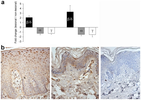
Figure 1. Overexpression of PPARβ/δ in psoriasis.
A. Fold-change of mRNA expression of PPARβ/δ (black columns), PPARα (shaded), and PPARγ (white) between lesional and non-lesional skin. Data shown represent mean ± s.d. from the GAIN dataset (left, n = 30) and the GSE14905 dataset (right, n = 28). p<10−3 for all data points shown. For each PPAR isoform, the probe yielding the highest hybridization signal was used to calculate the data shown (probesets 37152_at, 226978_at, 208510_at, respectively). B. Nuclear accumulation of PPARβ/δ in suprabasal epidermis in psoriasis skin lesions. Representative immunohistochemistry of paraffin-embedded lesional (left) and control (middle) skin samples stained with anti-PPARβ/δ, as well as staining control (right). Magnification 200×.

