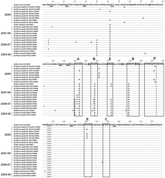
Figure 4. Amino acid comparison between HAl domains of H3N2 isolates and vaccine strains.
Dots represent amino acids similar to the consensus. The conserved amino acid residues at the receptor-binding site are shown as small rectangles. Alternative amino acids for sialic acid linkages of HA are underlined. The amino acid residues mapped at previously defined antigenic sites A–E are shown as large rectangles. The the potential N-glycosylation sites, with threshold value of >0.5, are double-underlined.

