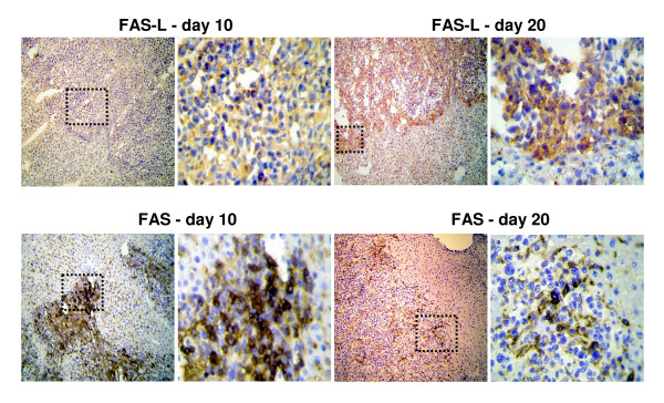
Figure 5.
(a) Immunohistochemistry: Representative images of FAS-L expression in liver metastases on day 10 (left) compared to increased FAS-L expression on day 20 (right). (b) Expression of FAS in liver metastasis on day 10 compared to day 20 demonstrating a decreased expression in the course of metastatic growth. DAB (3,3'-diaminobenzidine) brown color, Haemalaun blue color - nuclear counterstaining. Magnification ×100 and ×400. Case demonstrates area of magnification.

