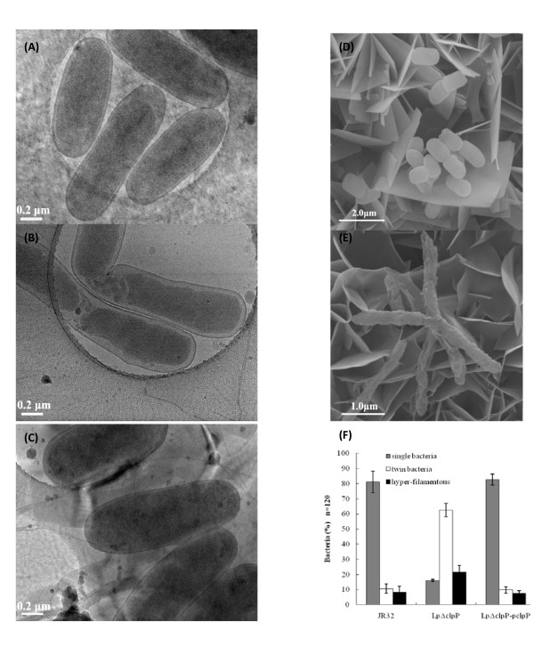
Figure 4.
Electron microscopy of stationary-phase L. pneumophila cells revealed cell elongation and abnormal division in the LpΔclpP mutant. Cyro-TEM of (A) JR32, (B) LpΔclpP and (C) LpΔclpP-pclpP and SEM of (D) JR32 and (E) LpΔclpP were carried out. Bar for (A), (B) and (C), 0.2 μm; Bar for (D), 2.0 μm; Bar for (E), 1.0 μm. (F) The percentages of normal and abnormal cells under cyro-TEM in the three L. pneumophila strains. Shown are the averages and standard deviations of three independent counts and the number of cells for each count is about 120 (n = 120).

