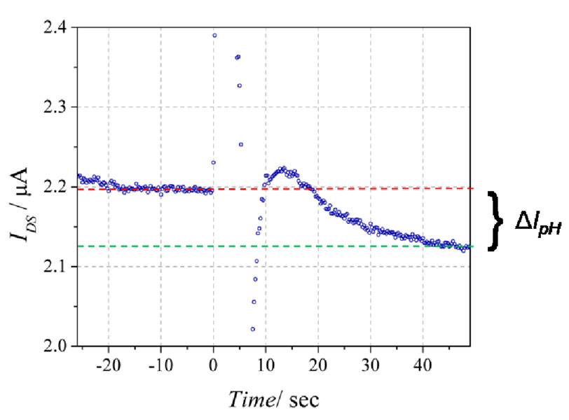
Figure 8.
Response [IDS(time)] of the sensor configured for IL-2 detection with 25 pg/mL IL-2 present during the protein-binding step (Fig. 1b-iii). At time = 0 the 100 µM urea solution was added to the pH 8.0 buffer. For this device, ΔIpH = 68.0 nA. The dashed red and green lines show the initial and final IDS levels, respectively.

