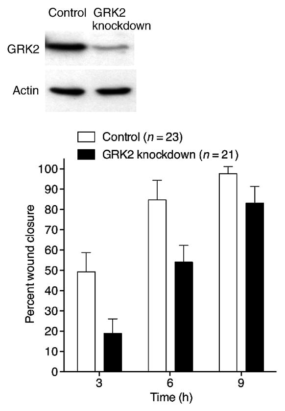
Figure 1. GRK2 positively regulates wound closure.

RNA interference-based silencing of GRK2 expression delays wound closure in MDCK cell monolayers. Western blots of whole-cell lysates from control and stable GRK2-knockdown cells (GRK2 knockdown) probed with anti-GRK2 and anti-actin antibodies are shown above the bar graph, demonstrating knockdown of GRK2. Mean percent wound closure values with standard errors of the mean (SEM) are shown at three times post-wounding for the indicated number of wounds from three independent experiments is shown in the graph. The smaller difference between the values for control and GRK2-knockdown cells at 9 h is due to the fact that wounds in the control monolayers had largely closed already by 9 h for the size of wounds made in these experiments.
