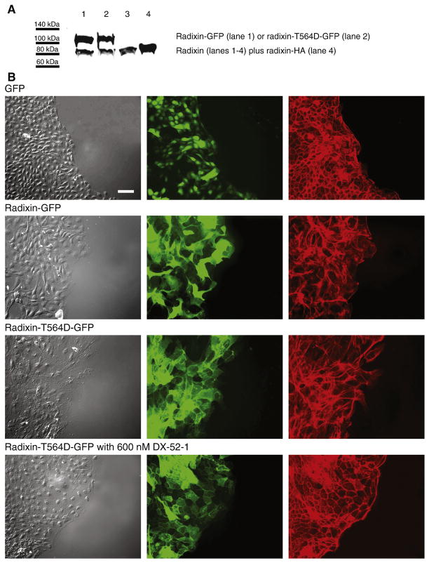
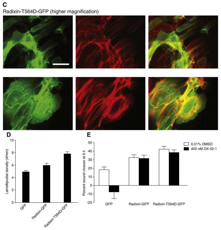
Figure 2. Expression of wild-type radixin and constitutively active radixin results in increased membrane protrusion and motility.
(A) Representative Western blot of whole-cell lysates from MDCK cells stably expressing radixin-GFP (lane 1), constitutively active, phosphomimetic radixin-T564D-GFP mutant (lane 2) or radixin-HA (lane 4; the radixin-HA cells were used in the experiment described in Figure 7) probed with anti-radixin antibody. Lane 3 consists of non-transfected MDCK cells, showing expression of endogenous radixin alone. The levels of expression were calculated from the intensity of bands on three separate Western blots as 222% for radixin-GFP, 137% for radixin-T564D-GFP and 112% for radixin-HA that of endogenous radixin. (B) Fluorescence images of wounded monolayers of cells expressing GFP alone, radixin-GFP or radixin-T564D-GFP in the presence or absence of the radixin inhibitor DX-52-1, as indicated. Confluent monolayers of MDCK cells stably expressing GFP alone, radixin-GFP or radixin-T564D-GFP were wounded and then fixed 6 h later and processed for imaging. From left to right, each row consists of a differential interference contrast image, a fluorescence image showing GFP fluorescence and a second fluorescence image showing TRITC-phalloidin staining of filamentous actin. All images in B are at the same magnification, with the scale bar corresponding to 50 μm. (C) Higher magnification fluorescence images of wounded sheets of cells expressing GFP alone or radixin-T564D-GFP 6 h after wounding. From left to right, each row consists of a fluorescence image showing GFP fluorescence, a second fluorescence image showing TRITC-phalloidin staining of filamentous actin and a merged image. All images in C are at the same magnification, with the scale bar corresponding to 50 μm. (D) Lamellipodial density at the wound margin (number of lamellipodial membrane protrusions at the wound edge divided by margin perimeter length) 6 h after wounding of monolayers of cells expressing GFP alone, radixin-GFP or radixin-T564D-GFP-expressing cells. Values are mean and SEM for 6 wounds from three independent experiments. (E) Percent wound closure at 6 h post-wounding in monolayers of cells expressing GFP alone, radixin-GFP and radixin-T564D-GFP in the presence or absence of DX-52-1 (mean and SEM for 12 wounds in each case from three independent experiments).


