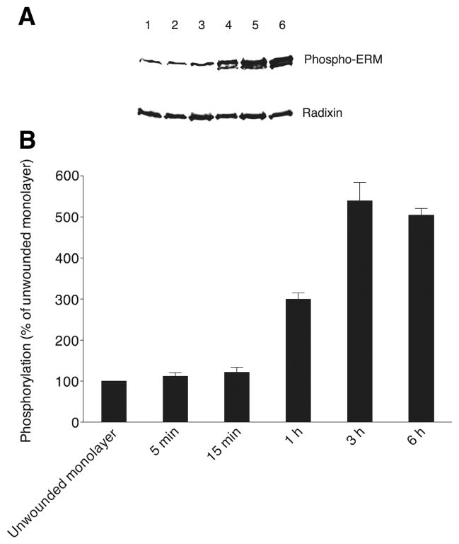
Figure 6. Phosphorylation of ERM proteins increases during the most active migratory phase of wound closure.
(A) Phospho-ERM levels are shown on a representative Western blot probed with anti-phospho-ERM antibody at the top for an unwounded monolayer (lane 1) and at the following times after wounding: 5 min (lane 2), 15 min (lane 3), 1 h (lane 4), 3 h (lane 5) and 6 h (lane 6). The same blot stripped and reprobed with anti-radixin antibody is shown at the bottom. (B) Levels (mean and SEM) of ERM phosphorylation as a function of time after wounding are shown, derived from band intensities on Western blots for three separate experiments as in A, with values expressed as a percent of the level of phospho-ERM in unwounded monolayers (normalization to the parallel unwounded monolayer for each experiment prior to statistics).

