
Fig. 6. Lipid accumulation in C2C12 myotubes treated with HIV-PIs.
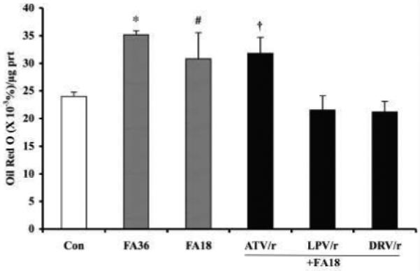
C2C12 myotubes were treated for 18 h in the presence of vehicle (DMSO) alone (Con) or with HIV-PIs in combination (ATV/r, LPV/r, and DRV/r; 20/5 μmol/l, respectively). The cells were then washed with HBSS and the Con cells were incubated with vehicle alone and the HIV-PI pretreated cells were incubated with 200/100 μmol/l oleate/palmitate for an additional 18 h. In some experiments, the myotubes were treated with only the fatty acids (FA) for 18 h (FA18) or 36 h (FA36). At the end of the incubation period, lipid in the cells was stained with 3% Oil Red O and quantitated as described in Methods section. The presented data are mean ± SEM of %Oil Red O stain/μg protein (n =3). (*.#FA groups significantly different from Con group, p < 0.0001 and p < 0.05, respectively).