
Fig. 3.
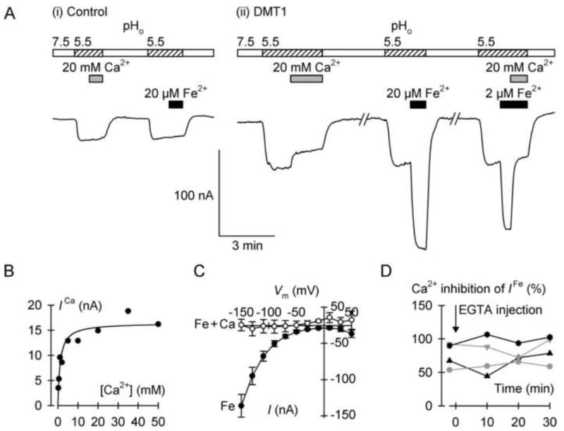
Calcium inhibition of the DMT1-mediated H+ leak and Fe2+-evoked currents. (A) Typical continuous current recordings at membrane potential (Vm) of 70mV in a control oocyte (i) and in an oocyte expressing DMT1 (ii). Oocytes were superfused with Ca2+-free medium at pH 7.5 for the periods shown by the empty bars, or with Ca2+-free medium at pH 5.5 (hatched bars). We presented 20 mM Ca2+ as indicated by the gray bars and 20 μM or 2 μM Fe2+ (black bars). (B) Ca2+-induced current (ICa) in the absence of Fe2+ metal ion as a function of Ca2+ concentration. Data were fit by equation 3 to describe the Ca2+ inhibition of the DMT1-mediated H+ leak current: Imax = 16.4 ± 1.0 nA, KiCa = 1.1 ± 0.3 mM (adjusted r2 = 0.88, P < 0.001). (C) Current-voltage relationships of the currents induced by 2 μM Fe2+ alone (Fe, filled symbols) or 2 μM Fe2+ plus 20 mM Ca2+ (Fe + Ca, empty symbols) (n = 4, paired). (D) Effect of intracellular Ca2+ chelation on the Ca2+ inhibition of the DMT1-mediated Fe2+-evoked current. We voltage-clamped at −70 mV four oocytes expressing DMT1, and measured currents evoked by 2 μM Fe2+ alone or 2 μM Fe2+ plus 20 mM Ca2+ before (−2 min) and after (10, 20, 30 min) microinjection with EGTA. Each symbol shape represents an individual oocyte. The mean Fe2+-evoked current at −2 min was −88 (SD 64) nA and did not change with time (one-way repeated measures ANOVA, P = 0.83). We obtained percent inhibition by Ca2+ and found no effects over time (one-way repeated measures ANOVA, P = 0.18).