Figure 8.
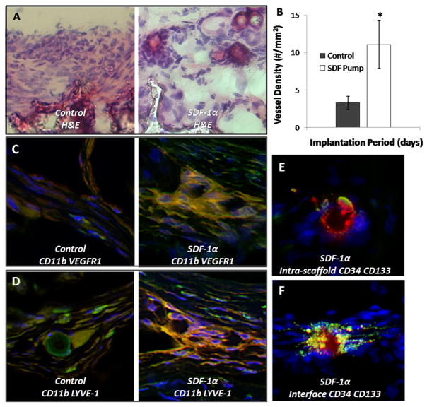
SDF-1α treatment improves EPC engraftment and participation of macrophage subsets in angiogenesis and lymphangiogenesis. H&E staining was used to examine the cellular response at the interface. Control scaffolds have a thick, dense cellular response (A) at the interface while SDF-1α group has a weaker cell response with the appearance of budding vessels at the interface. Quantification of vessel density within the scaffold implants reveals a significant difference in the degree of vessel formation (B), Student t-test (P < 0.05). Control scaffolds also have few cells co-expressing CD11b (green) and VEGFR1 (red) (C) in agreement with a low number of budding vessels. However, we do find the groups of cells expressing these markers in SDF-1α scaffolds. Control scaffolds have few cells staining positive for CD11b (green) LYVE-1 (red) (D) which are involved in lymphogenesis, while SDF-1α have organized groups of cells staining positive. The presence of EPC cells inside the scaffold (E) and at the interface (F) of SDF-1α modified scaffolds was monitored by expression of CD34+ (green) CD133+ (red) markers.

