
Figure 4. Realization of composite MS-laden scaffolds with sacrificial content.
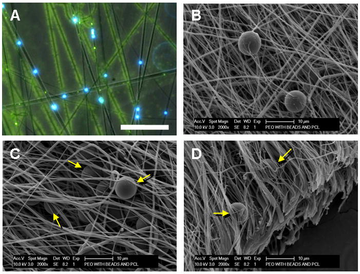
Bright-field with overlaid fluorescent image (A, 4X, Scale bar = 50 μm) and SEM (B, Scale bar = 20 μm) of PEO/PCL/MS composite. In (A), blue shows MS, green shows PCL fibers, and black shows sacrificial PEO fibers within the composite structure. After PEO removal, microspheres remain entrapped and distributed between the remaining PCL fibers (C and D, arrows, Scale bar = 10 μm).