
Fig. 1.
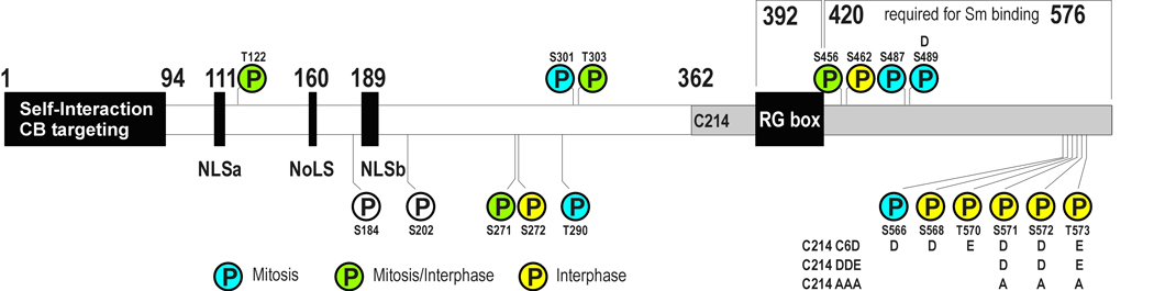
Schematic representation of the binding domains of coilin. Phosphoresidues identified by MS/MS are indicated. Residues identified from mitotic samples by MS/MS are colored blue, interphase samples are colored yellow, and residues found phosphorylated in both mitotic and interphase samples are green. S184 and S202 were identified by mutagenesis studies. Residues replaced in coilin mutants are indicated