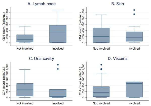
Figure 1.

Higher CD4 T-cell counts are associated with KS involvement of lymph nodes, but not other presentations, among HIV-infected children. Box plots display the CD4 T-cell count quartiles (minimum, 25th, 50th, and 75th percentile, and maximum) and outliers (dots) for those patients with and without KS lesions in a given location. CD4 T-cell values are shown for patients with and without KS involvement of the lymph nodes (panel A), skin (panel B), oral cavity (panel C), and viscera (panel D).
