Figure 4.

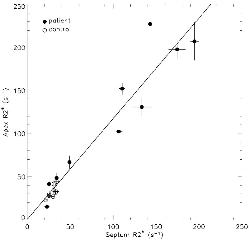
Correlation between septal and apical R2* of left ventricular cardiac wall. The solid line is the result of linear regression when all patients and control subjects are combined (r = 0.96, p < 0.001).

Correlation between septal and apical R2* of left ventricular cardiac wall. The solid line is the result of linear regression when all patients and control subjects are combined (r = 0.96, p < 0.001).