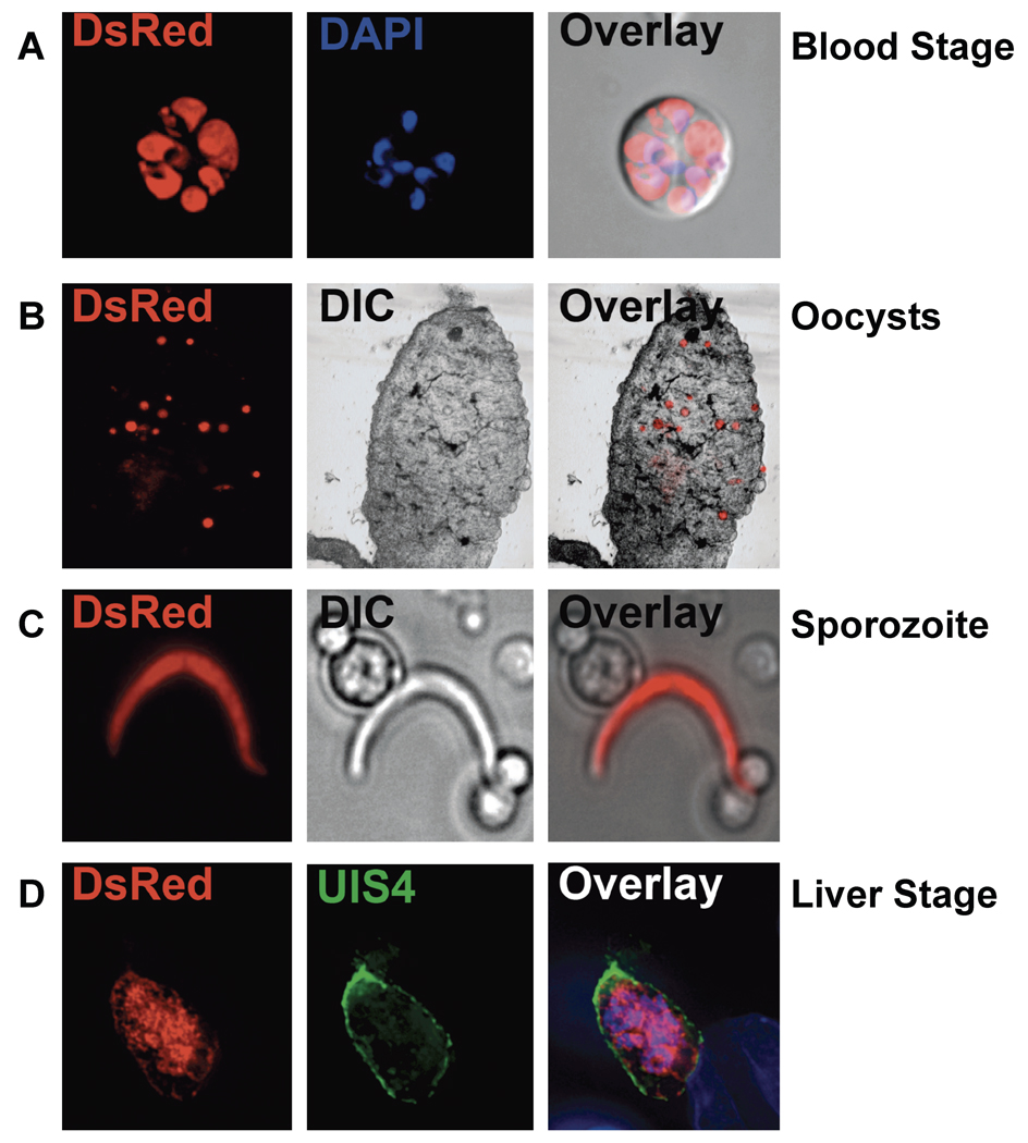
Figure 3.
Fluorescence microscopy of Pys1− parasites. Expression of DsRed in Pys1− parasites was evident in (A) live blood stages whose nuclei were visualized with 4',6-diamidino-2-phenylindole (DAPI) (Blue), (B) mosquito midgut oocysts in freshly dissected midguts, (C) the salivary gland sporozoite released from dissected mosquito salivary glands by grinding, and (D) liver stages in hepatoma cells fixed and visualized with an anti-UIS4 antibody and DAPI. Fluorescent and differential interference contrast (DIC) images were captured and merged (Overlay).

