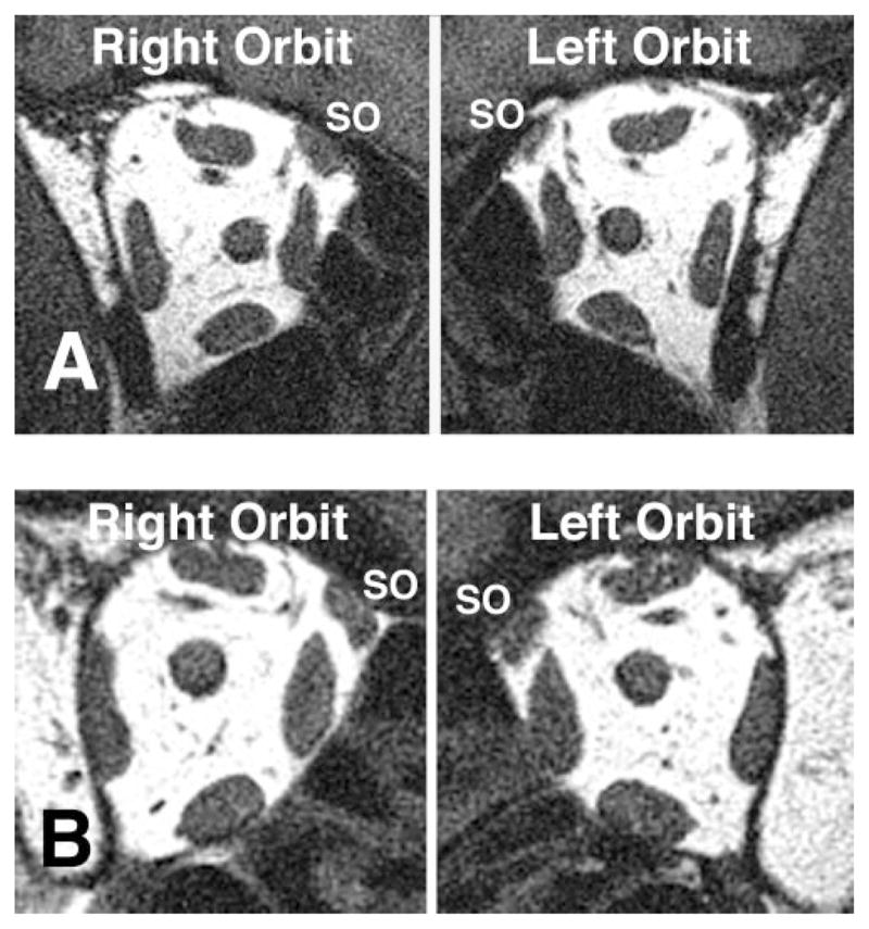
Figure 1.

Orbital MRI in quasi-coronal planes 8 mm posterior to the globe–optic nerve injection in central gaze in two subjects with clinically diagnosed SO palsy. (A) Subject 1 with left SO palsy and SO atrophy manifested by smaller left than right SO cross-sectional area. (B) Subject 2 with right SO palsy without SO atrophy.
