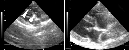
Fig. 6.
Echocardiographic evaluation of Case 2 before and after the heartworm removal (right parasternal short axis view, right outflow tract level) Left: before the procedure, many heartworms (arrowheads) are visible in the right ventricular outflow tract and pulmonary arteries. Right: after the procedure, no heartworms are visible in the right ventricular outflow tract and pulmonary arteries.

